Aortic Disease
Aortic Care Expert treatment for aortic conditions and cardiovascular health. Comprehensive Aortic Services Aortic aneurysm diagnosis and treatment Aortic dissection emergency care Aortic valve repair and replacement Advanced imaging and monitoring Minimally invasive procedures Expert Treatment Our specialized cardiac team provides comprehensive care for all aortic conditions, using advanced diagnostic tools and surgical techniques for optimal patient outcomes. Back to Blogs
Vital Signs
Vital Signs Understanding BP, pulse, temperature, and respiration for better health monitoring. Understanding Vital Signs Vital signs are the most basic measurements that indicate the status of your body’s essential functions. These measurements provide critical information about your overall health and help healthcare providers assess your condition quickly and effectively. The Four Primary Vital Signs Healthcare professionals monitor four key vital signs to assess your health status: 1. Blood Pressure (BP) Blood pressure measures the force of blood against your artery walls. It consists of two numbers: Systolic pressure (top number): Pressure when your heart beats Diastolic pressure (bottom number): Pressure when your heart rests between beats 2. Pulse Rate Your pulse rate indicates how many times your heart beats per minute. This measurement reflects your heart’s efficiency and overall cardiovascular health. 3. Body Temperature Body temperature indicates whether your body is fighting an infection or experiencing other health issues. Normal temperature can vary slightly throughout the day. 4. Respiration Rate Respiration rate measures how many breaths you take per minute. This vital sign helps assess your respiratory and overall health status. Normal Ranges for Adults Understanding normal ranges helps you recognize when your vital signs might indicate a health concern: Blood Pressure: Less than 120/80 mmHg (normal), 120-129/80 mmHg (elevated), 130/80 mmHg or higher (high) Pulse Rate: 60-100 beats per minute (bpm) at rest Body Temperature: 97-99°F (36.1-37.2°C) orally Respiration Rate: 12-20 breaths per minute at rest Factors That Affect Vital Signs Several factors can influence your vital signs, including: Age: Vital sign ranges vary by age group Activity level: Exercise and physical exertion affect all vital signs Stress and emotions: Anxiety and stress can elevate blood pressure and pulse Medications: Some medications can affect vital signs Time of day: Vital signs naturally fluctuate throughout the day Environmental factors: Temperature, altitude, and humidity can impact readings How to Measure Vital Signs at Home While professional medical equipment provides the most accurate readings, you can monitor some vital signs at home: Blood Pressure Monitoring Use a validated home blood pressure monitor Measure at the same time each day Sit quietly for 5 minutes before measuring Keep your arm at heart level Take multiple readings and average them Pulse Rate Measurement Place two fingers on your wrist or neck Count beats for 30 seconds and multiply by 2 Measure when you’re at rest Note any irregularities in rhythm Temperature Monitoring Use a digital thermometer for accuracy Wait 15 minutes after eating or drinking Take temperature orally, rectally, or under the arm Clean thermometer between uses When to Seek Medical Attention While some variations in vital signs are normal, certain readings require immediate medical attention: Blood Pressure Concerns Systolic pressure above 180 mmHg or diastolic above 110 mmHg Severe headache, chest pain, or shortness of breath with high BP Dizziness, confusion, or vision changes Pulse Rate Issues Resting pulse above 100 bpm or below 60 bpm Irregular or skipping heartbeats Pulse that’s difficult to feel or very weak Temperature Problems Fever above 103°F (39.4°C) Fever lasting more than 3 days Fever with severe headache, stiff neck, or rash Respiration Issues Breathing rate above 25 or below 12 breaths per minute Difficulty breathing or shortness of breath Labored breathing or chest pain with breathing Preventive Health Monitoring Regular monitoring of vital signs is an important part of preventive healthcare: Benefits of Regular Monitoring Early detection of health problems Better management of chronic conditions Improved communication with healthcare providers Peace of mind about your health status Recommended Monitoring Schedule Blood Pressure: Monthly for adults, more frequently if you have hypertension Pulse Rate: Weekly or when experiencing symptoms Temperature: When feeling unwell or experiencing fever symptoms Respiration: When experiencing breathing difficulties Why Choose Raksha Hospital for Vital Signs Monitoring At Raksha Hospital, we understand the importance of accurate vital signs assessment in providing quality healthcare. Our experienced medical team uses advanced monitoring equipment to ensure precise readings. Our Vital Signs Services Comprehensive vital signs assessment 24/7 monitoring for hospitalized patients Home monitoring equipment recommendations Education on self-monitoring techniques Interpretation of vital signs trends Back to Blogs
Heart Pumping Function
Heart Pumping Function Understanding ejection fraction and how to maintain optimal heart efficiency for a healthy cardiovascular system. Understanding Your Heart’s Pumping Function Your heart is a remarkable muscle that pumps blood throughout your body, delivering oxygen and nutrients to every cell. The efficiency of this pumping action is crucial for overall health and is measured by a key indicator called ejection fraction (EF). At Raksha Hospital, we help patients understand and optimize their heart function through comprehensive cardiac care. What is Ejection Fraction (EF)? Ejection fraction is a measurement that shows how much blood your heart pumps out with each beat. It’s expressed as a percentage and is one of the most important indicators of heart health. How EF is Calculated EF = (Amount of blood pumped out ÷ Total amount of blood in the heart) × 100 🟢 Normal EF: 50-70% Your heart is pumping efficiently and effectively. 🟡 Borderline EF: 41-49% Slightly below normal, may indicate early heart dysfunction. 🔴 Reduced EF: 40% or less Indicates heart failure or significant heart dysfunction. How Your Heart Pumps Blood Understanding the cardiac cycle helps explain how your heart maintains its pumping efficiency: 💓 Diastole (Filling Phase) Heart muscles relax Chambers fill with blood Valves open to allow blood flow Blood flows from atria to ventricles 💪 Systole (Pumping Phase) Heart muscles contract Blood is pumped out of ventricles Valves close to prevent backflow Blood flows to lungs and body The coordination between these phases determines your heart’s pumping efficiency and overall cardiac output. Factors That Affect Heart Pumping Function ✅ Positive Factors Regular Exercise: Strengthens heart muscle Healthy Diet: Provides essential nutrients Stress Management: Reduces heart strain Quality Sleep: Allows heart to rest and recover Regular Checkups: Early detection of issues ❌ Negative Factors High Blood Pressure: Strains heart muscle Smoking: Damages blood vessels Obesity: Increases heart workload Diabetes: Affects blood vessel health Excessive Alcohol: Weakens heart muscle How to Improve Your Heart’s Pumping Function 🏃♂️ Exercise and Physical Activity Regular cardiovascular exercise strengthens your heart muscle and improves its efficiency: Aerobic Exercise: Walking, cycling, swimming for 150 minutes/week Strength Training: 2-3 sessions per week to build muscle Interval Training: Alternating high and low intensity Consistency: Regular exercise is more important than intensity 🥗 Nutrition for Heart Health What you eat directly impacts your heart’s function and efficiency: Omega-3 Fatty Acids: Fish, nuts, seeds for heart muscle health Antioxidants: Fruits and vegetables to reduce inflammation Lean Proteins: Chicken, fish, legumes for muscle building Complex Carbohydrates: Whole grains for sustained energy Limit Salt: Reduce sodium to control blood pressure 🧘♀️ Lifestyle Modifications Daily habits that support optimal heart function: Stress Reduction: Meditation, yoga, deep breathing Quality Sleep: 7-9 hours per night for heart recovery Smoking Cessation: Eliminate tobacco use completely Moderate Alcohol: Limit to 1-2 drinks per day Weight Management: Maintain healthy BMI Medical Interventions for Heart Function When lifestyle changes aren’t enough, medical interventions can help improve heart pumping function: 💊 Medications ACE Inhibitors: Help heart work more efficiently Beta Blockers: Reduce heart rate and workload Diuretics: Remove excess fluid to reduce strain Blood Thinners: Prevent clots and improve circulation 🔬 Diagnostic Tests Echocardiogram: Visual assessment of heart function Cardiac MRI: Detailed heart structure and function Stress Test: Evaluate heart function under exertion Blood Tests: Check for underlying conditions ⚡ Advanced Treatments Cardiac Rehabilitation: Supervised exercise program Pacemaker: Regulate heart rhythm if needed Surgery: Repair or replace damaged heart valves Heart Transplant: Last resort for severe cases Monitoring Your Heart Function Regular monitoring helps track improvements and catch issues early: 📊 Regular Assessments Annual physical examinations Blood pressure monitoring Cholesterol level checks Blood sugar monitoring Weight and BMI tracking 🏥 Professional Monitoring Cardiologist consultations Regular echocardiograms Exercise stress tests Holter monitoring (24-hour ECG) Cardiac rehabilitation follow-ups Success Stories: Improving Heart Function Our cardiac care team has helped many patients improve their heart pumping function: “My EF was only 35% when I first came to Raksha Hospital. Through their cardiac rehabilitation program and lifestyle changes, I’ve improved to 55% in just 6 months. I feel stronger and more energetic than ever!” – Mr. Kumar, 62 “The personalized treatment plan and regular monitoring helped me understand my heart health better. My cardiologist explained everything clearly, and now I’m confident about managing my condition.” – Mrs. Devi, 58 Take Control of Your Heart Health Understanding your heart’s pumping function is the first step toward optimal cardiovascular health. Our expert cardiologists at Raksha Hospital are here to help you assess, improve, and maintain your heart function through comprehensive care and personalized treatment plans. Back to Blogs
Urinary Tract Infection (UTI)
Urinary Tract Infections (UTI) Recognize the signs early and get effective treatment for urinary tract infections. Expert urology care for optimal urinary health. Understanding Urinary Tract Infections (UTIs) Urinary tract infections are common bacterial infections that can affect any part of your urinary system, including the kidneys, bladder, ureters, and urethra. While UTIs are treatable, early recognition and proper treatment are crucial to prevent complications. At Raksha Hospital, our urology specialists provide comprehensive care for all types of urinary tract infections. What is a Urinary Tract Infection? A UTI occurs when bacteria enter the urinary tract and multiply, causing infection and inflammation. The most common type is a bladder infection (cystitis), but UTIs can also affect the kidneys (pyelonephritis) and urethra (urethritis). 🫁 Bladder Infection (Cystitis) The most common type of UTI, affecting the bladder. Usually caused by E. coli bacteria and more common in women. 🫀 Kidney Infection (Pyelonephritis) A more serious infection that can spread from the bladder to the kidneys. Requires immediate medical attention. 🚰 Urethra Infection (Urethritis) Infection of the urethra, the tube that carries urine from the bladder to outside the body. Common UTI Symptoms Recognizing UTI symptoms early is crucial for prompt treatment. Symptoms can vary depending on which part of the urinary tract is affected: 🚨 Common Symptoms Frequent Urination: Feeling the need to urinate more often than usual Burning Sensation: Pain or burning when urinating Urgency: Strong, persistent urge to urinate Cloudy Urine: Urine appears cloudy or has a strong odor Blood in Urine: Pink, red, or cola-colored urine ⚠️ Severe Symptoms (Seek Immediate Care) High Fever: Temperature above 101°F (38.3°C) Back Pain: Pain in the lower back or side Nausea & Vomiting: Gastrointestinal symptoms Confusion: Mental changes, especially in elderly Chills: Shaking or chills with fever Causes and Risk Factors Understanding what causes UTIs and who is at risk helps in prevention: 🔬 Common Causes Bacterial Entry: Bacteria from the digestive tract entering the urethra E. coli Bacteria: Most common cause, accounting for 80-90% of UTIs Sexual Activity: Can introduce bacteria into the urinary tract Catheter Use: Medical devices can introduce bacteria Blocked Urine Flow: Kidney stones or enlarged prostate ⚠️ Risk Factors Gender: Women are more prone due to shorter urethra Age: Elderly individuals have higher risk Diabetes: High blood sugar promotes bacterial growth Pregnancy: Hormonal changes affect urinary tract Menopause: Decreased estrogen levels Urinary Tract Abnormalities: Structural issues from birth Diagnosis of UTIs Accurate diagnosis is essential for effective treatment. Our urology specialists use various diagnostic methods: 🔬 Urine Analysis Examination of urine for white blood cells, red blood cells, and bacteria Dipstick test for nitrites and leukocyte esterase Microscopic examination of urine sediment 🧪 Urine Culture Growing bacteria from urine sample to identify specific organisms Determining which antibiotics are most effective Helping guide treatment decisions 📷 Imaging Tests Ultrasound to check for kidney stones or blockages CT scan for detailed urinary tract imaging X-rays for structural abnormalities 🔍 Cystoscopy Direct visualization of bladder and urethra Useful for recurrent UTIs or unusual symptoms Can detect structural problems Treatment Options for UTIs Treatment depends on the type and severity of the UTI. Our urology team provides personalized treatment plans: 💊 Antibiotic Treatment Antibiotics are the mainstay of UTI treatment: Common Antibiotics: Trimethoprim-sulfamethoxazole, nitrofurantoin, fosfomycin Duration: 3-7 days for simple UTIs, longer for complicated cases Effectiveness: Most UTIs respond well to appropriate antibiotics Follow-up: Important to complete the full course 🏥 Hospital Treatment Severe cases may require hospitalization: Intravenous Antibiotics: For severe kidney infections Fluid Therapy: To maintain hydration and kidney function Pain Management: To control severe symptoms Monitoring: Close observation of vital signs 🔧 Surgical Interventions Surgery may be needed for underlying structural issues: Kidney Stone Removal: If stones are causing blockages Prostate Surgery: For men with enlarged prostate Urinary Tract Repair: For structural abnormalities Prevention Strategies Preventing UTIs is often possible with simple lifestyle changes and good hygiene practices: 💧 Hydration and Urination Drink Plenty of Water: Aim for 6-8 glasses daily Urinate Frequently: Don’t hold urine for long periods Empty Bladder Completely: Take time to fully void Urinate After Sex: Helps flush out bacteria 🧼 Hygiene Practices Wipe Front to Back: Prevents bacteria from entering urethra Clean Genital Area: Gentle cleaning with mild soap Change Underwear Daily: Keep area clean and dry Avoid Irritating Products: Skip harsh soaps and douches 🥗 Diet and Lifestyle Cranberry Products: May help prevent UTIs Probiotics: Support healthy bacteria balance Avoid Irritants: Limit caffeine, alcohol, spicy foods Regular Exercise: Promotes overall health When to Seek Medical Care While some mild UTIs may resolve on their own, certain situations require immediate medical attention: 🚨 Seek Immediate Care High fever (above 101°F/38.3°C) Severe back or side pain Blood in urine Nausea and vomiting Confusion or mental changes Symptoms lasting more than 3 days 📞 Schedule Appointment Mild symptoms that don’t improve Recurrent UTIs (more than 2-3 per year) UTI during pregnancy UTI with diabetes or other chronic conditions Unusual symptoms or concerns Success Stories: Overcoming UTIs Our urology team has helped many patients successfully treat and prevent UTIs: “I suffered from recurrent UTIs for years. The team at Raksha Hospital identified the underlying cause and provided a comprehensive treatment plan. Now I’m UTI-free and feel much better!” – Mrs. Priya, 45 “The preventive strategies they taught me have been life-changing. I haven’t had a UTI in over a year, and I feel more confident about my urinary health.” – Ms. Anjali, 32 Get Expert UTI Care at Raksha Hospital Don’t let urinary tract infections affect your quality of life. Our experienced urology specialists provide comprehensive diagnosis, treatment, and prevention strategies for all types of UTIs. Early treatment can prevent complications and ensure a speedy recovery. Back to Blogs
Cardiac Care
Arrhythmia Understanding irregular heartbeats and their impact on your health. What is Arrhythmia? Arrhythmia is a condition where your heart beats in an irregular pattern – either too fast, too slow, or with an irregular rhythm. This can affect how well your heart pumps blood to the rest of your body. The heart normally beats in a coordinated, rhythmic pattern controlled by electrical signals, but when these signals become disrupted, arrhythmias occur. Understanding Normal Heart Rhythm The heart’s electrical system normally works like a sophisticated conductor: Sinoatrial (SA) Node: The heart’s natural pacemaker, located in the right atrium Atrioventricular (AV) Node: Acts as a relay station between atria and ventricles Bundle of His: Conducts electrical signals to the ventricles Purkinje Fibers: Distribute signals throughout the ventricular muscle Types of Arrhythmia Arrhythmias are classified based on their location and effect on heart rate: 1. Tachycardia (Fast Heart Rate) Supraventricular Tachycardia (SVT): Fast heart rate originating above the ventricles Atrial Fibrillation (AFib): Irregular and rapid heart rhythm in upper chambers Atrial Flutter: Regular but rapid atrial rhythm Ventricular Tachycardia (VT): Fast rhythm originating in the ventricles Wolff-Parkinson-White Syndrome: Extra electrical pathway causing rapid heartbeats 2. Bradycardia (Slow Heart Rate) Sick Sinus Syndrome: SA node dysfunction causing slow heart rate Heart Block: Impaired electrical conduction between atria and ventricles Bradycardia-Tachycardia Syndrome: Alternating slow and fast heart rates 3. Irregular Heart Rhythms Premature Beats: Extra or early heartbeats (PACs, PVCs) Atrial Fibrillation: Chaotic, irregular atrial rhythm Ventricular Fibrillation (VFib): Life-threatening chaotic ventricular rhythm Long QT Syndrome: Genetic condition causing dangerous arrhythmias Causes and Risk Factors Arrhythmias can develop from various causes: Primary Heart Conditions Coronary Artery Disease: Reduced blood flow damages heart muscle Heart Attack: Scar tissue disrupts electrical pathways Heart Failure: Enlarged heart chambers affect electrical conduction Heart Valve Disease: Structural changes affect heart function Cardiomyopathy: Heart muscle disease affecting electrical system Systemic Conditions High Blood Pressure: Strains heart muscle and electrical system Diabetes: Affects blood vessels and heart function Thyroid Disease: Hyperthyroidism can cause rapid heart rhythms Sleep Apnea: Oxygen deprivation affects heart rhythm Electrolyte Imbalances: Low potassium, magnesium, or calcium levels Lifestyle and Environmental Factors Smoking: Damages blood vessels and heart tissue Excessive Alcohol: “Holiday heart syndrome” and chronic damage Stress and Anxiety: Activates fight-or-flight response Certain Medications: Some drugs can cause arrhythmias Illegal Drugs: Cocaine, amphetamines, and others Symptoms and Clinical Presentation Arrhythmia symptoms can vary widely depending on the type and severity: Common Symptoms Palpitations: Feeling of skipped heartbeats, fluttering, or racing heart Chest Discomfort: Pain, pressure, or tightness in the chest Shortness of Breath: Difficulty breathing, especially with exertion Dizziness: Lightheadedness or feeling faint Fatigue: Unusual tiredness or weakness Fainting (Syncope): Loss of consciousness due to reduced blood flow Severe Symptoms (Medical Emergency) Chest Pain: Severe, crushing chest pain Severe Shortness of Breath: Difficulty breathing even at rest Loss of Consciousness: Fainting or near-fainting episodes Cardiac Arrest: No pulse or breathing Diagnosis and Testing Accurate diagnosis requires a comprehensive evaluation: Initial Assessment Medical History: Review of symptoms, risk factors, and family history Physical Examination: Heart sounds, pulse assessment, blood pressure Electrocardiogram (ECG/EKG): Records heart’s electrical activity Advanced Diagnostic Tests Holter Monitor: 24-48 hour continuous ECG recording Event Monitor: Records heart rhythm during symptoms Echocardiogram: Ultrasound imaging of heart structure Stress Test: Evaluates heart rhythm during exercise Electrophysiology Study (EPS): Invasive test to map electrical pathways Cardiac MRI: Detailed imaging of heart tissue Treatment Strategies Treatment depends on the type, severity, and underlying cause of the arrhythmia: 1. Lifestyle Modifications Heart-Healthy Diet: Low-sodium, balanced nutrition Regular Exercise: Supervised cardiac rehabilitation Stress Management: Meditation, yoga, counseling Smoking Cessation: Complete tobacco avoidance Alcohol Moderation: Limit or eliminate alcohol consumption Sleep Hygiene: Treat sleep apnea and ensure quality sleep 2. Medications Antiarrhythmic Drugs: Control heart rhythm and rate Beta-Blockers: Slow heart rate and reduce blood pressure Calcium Channel Blockers: Control heart rate and rhythm Anticoagulants: Prevent blood clots in atrial fibrillation Electrolyte Supplements: Correct potassium, magnesium deficiencies 3. Medical Procedures Cardioversion: Electrical shock to restore normal rhythm Catheter Ablation: Destroy abnormal electrical pathways Pacemaker Implantation: For slow heart rates Implantable Cardioverter Defibrillator (ICD): For dangerous arrhythmias Maze Procedure: Surgical treatment for atrial fibrillation Prevention and Risk Reduction While some arrhythmias can’t be prevented, many risk factors can be managed: Regular Health Screenings: Early detection of underlying conditions Blood Pressure Control: Maintain healthy levels Diabetes Management: Keep blood sugar stable Cholesterol Control: Maintain healthy lipid levels Healthy Lifestyle: Balanced diet, exercise, stress management Medication Review: Regular assessment of drug interactions Genetic Counseling: For families with inherited conditions Living with Arrhythmia With proper management, most people with arrhythmias can lead active, fulfilling lives: Regular Monitoring: Frequent follow-up with healthcare providers Medication Adherence: Take medications exactly as prescribed Symptom Awareness: Recognize and report changes promptly Emergency Planning: Know when and how to seek urgent care Support Systems: Family, friends, and support groups Lifestyle Adjustments: Adapt activities to energy levels When to Seek Emergency Care Seek immediate medical attention for these symptoms: Chest pain lasting more than 5 minutes Severe shortness of breath Fainting or loss of consciousness Rapid, irregular heartbeat with dizziness Heart rate over 150 or under 50 beats per minute Severe weakness or confusion Back to Blogs
Urethral Stricture
Understanding Urethral Stricture Comprehensive guide to urethral narrowing: causes, symptoms, diagnosis, and advanced treatment options. What is Urethral Stricture? A urethral stricture is a narrowing of the urethra, the tube that carries urine from the bladder out of the body. This condition can affect both men and women, though it’s more common in men due to the longer length of the male urethra. The narrowing can occur anywhere along the urethra and can range from mild to severe, significantly impacting urinary function and quality of life. Anatomy of the Urethra The urethra is a muscular tube that serves as the final pathway for urine elimination. In men, it also carries semen during ejaculation. The urethra has several segments: Prostatic urethra: Passes through the prostate gland Membranous urethra: Short segment through the pelvic floor muscles Penile urethra: Runs through the penis Bulbar urethra: Located in the bulb of the penis Causes of Urethral Stricture Understanding the underlying causes helps in prevention and guides treatment decisions. Urethral strictures can develop from various factors: Trauma and Injury Pelvic fractures: Can damage the urethra during accidents Penetrating injuries: Gunshot wounds, stab wounds, or other penetrating trauma Catheter trauma: Prolonged or improper catheter use Surgical procedures: Complications from urological or gynecological surgeries Infections and Inflammation Sexually transmitted infections: Gonorrhea, chlamydia, and other STIs Urinary tract infections: Recurrent UTIs can cause scarring Prostatitis: Inflammation of the prostate gland Urethritis: Inflammation of the urethra itself Medical Conditions Benign prostatic hyperplasia (BPH): Enlarged prostate can compress the urethra Urethral cancer: Rare but can cause strictures Radiation therapy: Can cause scarring in the pelvic area Autoimmune conditions: Some inflammatory diseases affect the urethra Congenital Factors Some individuals are born with urethral abnormalities that predispose them to strictures later in life. These may include: Narrow urethra from birth Urethral valves or other structural abnormalities Hypospadias or epispadias Symptoms of Urethral Stricture The symptoms of urethral stricture can vary depending on the severity and location of the narrowing. Early recognition of these symptoms is crucial for timely treatment: Urinary Symptoms Weak urine stream: Reduced force and flow of urination Urinary spraying: Urine may spray in multiple directions Straining to urinate: Need to push or strain to start urination Incomplete emptying: Feeling that the bladder isn’t fully empty Frequent urination: Need to urinate more often than usual Urgency: Sudden, strong need to urinate Complications and Advanced Symptoms Recurrent urinary tract infections: Due to incomplete bladder emptying Bladder stones: Can form due to urinary stasis Urinary retention: Complete inability to urinate (medical emergency) Hydronephrosis: Swelling of the kidneys due to urine backup Kidney damage: Long-term complications if left untreated Pain and Discomfort Pain or burning during urination Lower abdominal or pelvic pain Pain in the penis or perineum Discomfort during sexual activity Diagnosis of Urethral Stricture Accurate diagnosis is essential for determining the best treatment approach. Your urologist will use several diagnostic methods: Medical History and Physical Examination Detailed discussion of symptoms and their progression History of trauma, infections, or surgeries Physical examination of the genital area Digital rectal examination to assess the prostate Diagnostic Tests Uroflowmetry: Measures the speed and volume of urine flow Post-void residual: Measures urine remaining in bladder after urination Urethroscopy: Direct visualization of the urethra using a small camera Retrograde urethrogram: X-ray imaging with contrast dye Voiding cystourethrogram: X-ray during urination Ultrasound: To assess bladder and kidney function Treatment Options for Urethral Stricture Treatment choice depends on the stricture’s location, length, severity, and whether it’s a first-time occurrence or a recurrence. Your urologist will recommend the most appropriate option: Minimally Invasive Procedures Urethral Dilation Urethral dilation involves gradually stretching the narrowed area using progressively larger dilators. This is often used for: Short, simple strictures First-time strictures Patients who prefer non-surgical options Pros: Minimally invasive, quick recovery, outpatient procedure Cons: Higher recurrence rate, may need repeated procedures Internal Urethrotomy This procedure uses a special instrument to cut the stricture from inside the urethra. It’s suitable for: Short strictures (less than 1 cm) Bulbar urethra strictures Patients who want to avoid open surgery Pros: Less invasive than open surgery, shorter recovery time Cons: Moderate recurrence rate, may require multiple procedures Surgical Treatment Urethroplasty Urethroplasty is the gold standard for treating urethral strictures, especially for longer or recurrent strictures. This surgical procedure involves: Removing the scarred, narrowed section Reconstructing the urethra using healthy tissue Various techniques depending on stricture characteristics Types of Urethroplasty Excision and primary anastomosis: For short strictures, removes the narrowed segment and reconnects the healthy ends Graft urethroplasty: Uses tissue from the mouth (buccal mucosa) or other areas to widen the urethra Flap urethroplasty: Uses local skin flaps to reconstruct the urethra Two-stage urethroplasty: For complex cases, involves creating a temporary opening and later reconstructing the urethra Laser Treatment Laser therapy uses focused light energy to remove scar tissue and open the stricture. It’s particularly useful for: Short, simple strictures Patients who prefer laser treatment Cases where traditional methods have failed Stent Placement Urethral stents are temporary or permanent devices that hold the urethra open. They’re used for: Patients who cannot undergo surgery Temporary relief while planning definitive treatment Palliative care in certain situations Recovery and Follow-up Care Proper recovery and follow-up are crucial for successful treatment outcomes: Post-Procedure Care Catheter care: Proper care of urinary catheter if placed Medications: Antibiotics and pain management as prescribed Activity restrictions: Avoid heavy lifting and strenuous activity Hygiene: Keep the area clean and dry Long-term Follow-up Regular check-ups with your urologist Monitoring for recurrence of symptoms Uroflowmetry tests to assess urinary function Lifestyle modifications to prevent recurrence Prevention Strategies While not all urethral strictures can be prevented, certain measures can reduce your risk: Lifestyle Modifications Safe sex practices: Use protection to prevent STIs Proper catheter care: If you need a catheter, follow care instructions carefully Stay hydrated: Drink plenty of water to maintain urinary health Regular exercise: Maintain overall health and circulation Medical Prevention Prompt treatment of urinary tract infections Regular check-ups if you have risk factors Proper management of underlying medical conditions Seek immediate care for urinary symptoms Why Choose Raksha Hospital for Urethral Stricture Treatment At Raksha Hospital, we understand the impact that urethral strictures can have on your quality of life. Our experienced urology team provides comprehensive care using the latest diagnostic and treatment techniques.
Arrhythmia

Heart Health Small daily habits make a big difference. Five pillars Don’t smoke; limit alcohol 30–45 min activity most days Eat more plants, fiber, and healthy fats Sleep 7–8 hours Manage stress Screening Check BP, sugar, and lipids at recommended intervals — especially if you have family history or are over 40. Back to Blogs
Cardiomyopathy
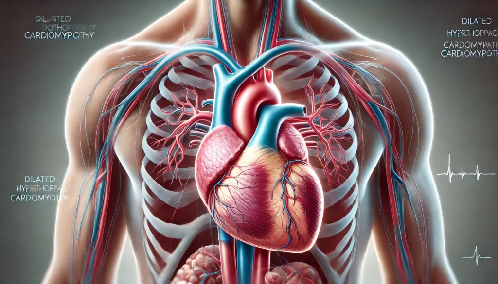
Cardiomyopathy Expert treatment for heart muscle disease and cardiac conditions. Cardiomyopathy Treatment Comprehensive heart muscle disease diagnosis Advanced cardiac imaging and testing Personalized treatment plans Medication management and monitoring Lifestyle counseling and support Expert Care Our specialized cardiologists provide comprehensive care for all types of cardiomyopathy, using advanced diagnostic tools and personalized treatment approaches for optimal outcomes. Back to Blogs
Coronary Artery Disease
Coronary Artery Disease Understanding plaque buildup in heart arteries and preventing life-threatening complications through early detection and treatment. What is Coronary Artery Disease (CAD)? Coronary Artery Disease (CAD) is the most common type of heart disease and a leading cause of death worldwide. It occurs when the coronary arteries, which supply blood to the heart muscle, become narrowed or blocked due to the buildup of fatty deposits called plaque. This process, known as atherosclerosis, can lead to chest pain (angina), heart attacks, and other serious complications. How Coronary Artery Disease Develops Understanding the development of CAD helps in prevention and early intervention: 🔬 Stage 1: Initial Damage Damage to the inner lining of coronary arteries, often caused by: High blood pressure High cholesterol levels Smoking and tobacco use Diabetes and insulin resistance Inflammation in the body 🩸 Stage 2: Plaque Formation Cholesterol and other substances accumulate at the damaged site: LDL cholesterol penetrates the artery wall White blood cells try to repair the damage Plaque begins to form and grow Artery narrows, reducing blood flow ⚠️ Stage 3: Complications As plaque continues to build up: Arteries become significantly narrowed Blood flow to heart muscle is reduced Plaque can rupture, causing blood clots Complete blockage leads to heart attack Risk Factors for Coronary Artery Disease Several factors increase your risk of developing CAD. Some can be controlled, while others cannot: ✅ Controllable Risk Factors High Blood Pressure: Damages artery walls over time High Cholesterol: LDL cholesterol contributes to plaque formation Smoking: Damages blood vessels and reduces oxygen Diabetes: High blood sugar damages arteries Obesity: Increases strain on heart and blood vessels Physical Inactivity: Weakens heart muscle and circulation Poor Diet: High in saturated fats, trans fats, and sodium Excessive Alcohol: Can raise blood pressure and cholesterol ❌ Uncontrollable Risk Factors Age: Risk increases with age, especially after 45 for men and 55 for women Gender: Men have higher risk, but women’s risk increases after menopause Family History: Genetic predisposition to heart disease Ethnicity: Higher risk in certain ethnic groups Previous Heart Disease: History of heart attack or stroke Common Symptoms of Coronary Artery Disease CAD symptoms can vary widely, and some people may not experience any symptoms until the disease is advanced: 🚨 Common Symptoms Chest Pain (Angina): Pressure, tightness, or burning sensation Shortness of Breath: Difficulty breathing during physical activity Fatigue: Unusual tiredness, especially during exertion Dizziness: Lightheadedness or feeling faint Nausea: Feeling sick to your stomach ⚠️ Severe Symptoms (Emergency) Severe Chest Pain: Crushing pain that doesn’t go away Pain Spreading: To arms, neck, jaw, or back Cold Sweats: Profuse sweating with chest discomfort Shortness of Breath: Severe difficulty breathing Nausea and Vomiting: Accompanied by chest pain 🤐 Silent CAD Some people experience no symptoms, especially those with diabetes or elderly individuals. Regular checkups are crucial for early detection. Diagnosing Coronary Artery Disease Early diagnosis is crucial for effective treatment. Our cardiologists use various diagnostic methods: 🔬 Blood Tests Cholesterol Panel: Measures total, LDL, HDL cholesterol and triglycerides Cardiac Enzymes: Detect heart muscle damage Inflammatory Markers: C-reactive protein (CRP) levels Blood Sugar: Check for diabetes or prediabetes 📷 Imaging Tests Electrocardiogram (ECG): Records heart’s electrical activity Echocardiogram: Ultrasound of heart structure and function Stress Test: Monitor heart during exercise Cardiac CT Scan: Detailed images of heart and arteries Cardiac MRI: High-resolution heart imaging 🔍 Invasive Procedures Coronary Angiogram: X-ray with contrast dye to see arteries Cardiac Catheterization: Direct examination of heart chambers Intravascular Ultrasound: Detailed artery wall imaging Treatment Options for Coronary Artery Disease Treatment depends on the severity of CAD and may include lifestyle changes, medications, and procedures: 💊 Medications Various medications help manage CAD: Statins: Lower cholesterol and reduce inflammation Beta Blockers: Reduce heart rate and blood pressure ACE Inhibitors: Relax blood vessels and lower blood pressure Calcium Channel Blockers: Relax heart muscle and blood vessels Nitroglycerin: Relieve chest pain during angina attacks Blood Thinners: Prevent blood clots and reduce heart attack risk 🔧 Medical Procedures When medications aren’t enough: Angioplasty: Open blocked arteries with a balloon Stent Placement: Keep arteries open with mesh tubes Coronary Bypass Surgery: Create new blood flow routes Atherectomy: Remove plaque from arteries 🏥 Cardiac Rehabilitation Comprehensive program for recovery: Supervised Exercise: Safe physical activity program Education: Learn about heart-healthy living Counseling: Emotional support and stress management Lifestyle Changes: Diet, smoking cessation, weight management Preventing Coronary Artery Disease Prevention is the best approach to CAD. Many risk factors can be controlled through lifestyle changes: 🥗 Heart-Healthy Diet Mediterranean Diet: Rich in fruits, vegetables, whole grains, fish Limit Saturated Fats: Choose lean proteins and low-fat dairy Reduce Sodium: Aim for less than 2,300mg per day Increase Fiber: 25-30 grams daily from whole foods Omega-3 Fatty Acids: Found in fatty fish, nuts, and seeds 🏃♂️ Regular Exercise Aerobic Activity: 150 minutes of moderate exercise weekly Strength Training: 2-3 sessions per week Start Slowly: Gradually increase intensity and duration Stay Active: Find activities you enjoy and can maintain 🚭 Lifestyle Modifications Quit Smoking: Seek help for smoking cessation Limit Alcohol: Moderate consumption (1-2 drinks daily) Manage Stress: Practice relaxation techniques Maintain Healthy Weight: BMI between 18.5-24.9 Regular Checkups: Monitor blood pressure, cholesterol, and blood sugar When to Seek Emergency Medical Care Recognizing the signs of a heart attack can save your life. Seek immediate medical attention if you experience: 🚨 Heart Attack Warning Signs Chest Pain: Pressure, squeezing, or fullness that lasts more than a few minutes Pain Spreading: To arms, neck, jaw, or back Shortness of Breath: With or without chest discomfort Cold Sweats: Profuse sweating Nausea: Feeling sick to your stomach Lightheadedness: Feeling faint or dizzy Remember: Don’t wait more than 5 minutes to call emergency services if you suspect a heart attack. Success Stories: Overcoming Coronary Artery Disease Our cardiac care team has helped many patients manage CAD and improve their heart health: “After my heart attack, I was scared and didn’t know how to manage my condition. The team at Raksha Hospital created a comprehensive treatment plan that included medication, lifestyle changes, and cardiac rehabilitation. Now I feel stronger and more confident about my heart health.” – Mr. Reddy, 58 “The preventive care program helped me understand my risk factors and make necessary changes. Through diet, exercise, and medication, my cholesterol and blood pressure are now under control, and I feel much better.” – Mrs. Iyer, 62 Take Control of Your Heart Health Coronary Artery Disease is serious but manageable
Echocardiogram
Echocardiogram Test Advanced heart ultrasound testing for accurate cardiac diagnosis. What is an Echocardiogram? An echocardiogram is a safe, non-invasive ultrasound test that uses high-frequency sound waves to create detailed images of your heart. This diagnostic tool provides cardiologists with real-time information about your heart’s structure, function, and blood flow. Types of Echocardiograms Transthoracic Echo (TTE): Standard heart ultrasound – most common type Transesophageal Echo (TEE): Detailed imaging through esophagus Stress Echo: Heart function assessment during exercise 3D Echo: Advanced three-dimensional imaging What It Shows Echocardiogram provides comprehensive information about heart function, valve problems, structural abnormalities, and blood flow patterns. It’s essential for diagnosing heart conditions and monitoring treatment effectiveness. Back to Blogs